Login
SubscribeADVERTISEMENT


Visualizing Axon Pruning
Tiffany Garbutt, PhD | Oct 2, 2023 | 2 min read
During development, neurons trim hundreds of excess axons in an intricately coordinated destructive process.

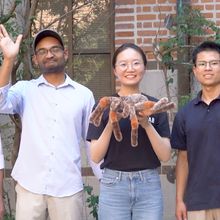
A Big Data Approach to Life Science
Mariella Bodemeier Loayza Careaga, PhD | Oct 2, 2023 | 2 min read
As a group leader at the Broad Institute, Shantanu Singh develops tools to tackle high-dimensional biological data.

Turning the PAGE: Tips for Protein Electrophoresis and Western Blotting
The Scientist’s Creative Services Team | 1 min read
In this webinar, Kelly Wolfe will discuss the dos and don’ts of protein electrophoresis and western blotting.

Striking a Balance for Perfect Images
The Scientist Staff | Oct 2, 2023 | 2 min read
Advanced microscope systems enable researchers to perform high-resolution live-cell imaging, while maintaining cellular health.

A Picture Sparks a Thousand Words
Meenakshi Prabhune, PhD | Oct 2, 2023 | 2 min read
A scientific image can conceal even more than it reveals. Scientists can now share their untold behind-the-image stories in our new Science Snapshot column.

BD CellView™ Image Technology: Sort What You See
BD Biosciences | 1 min read
Camera-free imaging unlocks new cell sorting applications.

Emerging from Silence: Capturing the First Heartbeat
Danielle Gerhard, PhD | Sep 27, 2023 | 5 min read
In the developing zebrafish, a noisy and asynchronous activity jumpstarts the heart’s journey to coordinated beating.

Assembloids Unlock the Roles of Key Neurodevelopment Disease Genes
Aparna Nathan, PhD | Sep 27, 2023 | 3 min read
Brain-like tissue grown in a dish mimics critical periods for development and reveals how it can go wrong.

Modeling Human Disease and Development with Organoids
The Scientist’s Creative Services Team | 1 min read
Discover how scientists use cardiac and skin organoids to study differentiation and toxicity.

Niche Interactions Lock Down Leukemia Cells
Deanna MacNeil, PhD | Sep 24, 2023 | 3 min read
Researchers unravel the mystery of epithelial to mesenchymal transition in acute lymphoblastic leukemia with co-culture techniques, CRISPR-screening, and RNA sequencing.

Piet Borst Wins a Lasker Award for Scientific Excellence
Laura Tran, PhD | Sep 21, 2023 | 7 min read
This year's Lasker~Koshland Award for Special Achievement was awarded to Piet Borst for his stellar work on cell organelles, trypanosomes, and cancer drug resistance.

Discover the Freshness of Frozen PBMCs
The Scientist’s Creative Services Team | 1 min read
Researchers discuss how peripheral blood mononuclear cell (PBMC) cryopreservation affects T cell activation and immune cell phenotypes.
2023 Ig Nobel Prize for Gripping Work on Dead Spiders
Meenakshi Prabhune, PhD | Sep 15, 2023 | 3 min read
Rice University researchers claimed the Ig Nobel Prize for upleveling biorobotics by transforming deceased spiders into robotic grippers.

Ian Wilmut, Famed Scientist Who Led the Creation of Dolly the Sheep, Died at 79
Shelby Bradford, PhD | Sep 12, 2023 | 3 min read
Knighted in 2008, Sir Ian Wilmut revolutionized the field of cloning, stem cell research, and regenerative medicine.

Introducing GMP-Grade Recombinant Cytokines for Stem Cell Research
The Scientist’s Creative Services Team and Sino Biological | 3 min read
Cutting edge technology delivers cytokines with high purity, high bioactivity, high batch-to-batch consistency, and high stability.

Hair Turns Gray Due to Stuck Stem Cells
Mariella Bodemeier Loayza Careaga, PhD | Sep 8, 2023 | 3 min read
Hair-coloring stem cells must swing back and forth between their maturity states to give hair its color.

Magnifying Curiosity with a Pocket Microscope
Danielle Gerhard, PhD | Sep 8, 2023 | 5 min read
Microscopes were inaccessible to most of the world until Manu Prakash and Jim Cybulski put their engineering prowess to the test.

Turning Organoids Inside Out
The Scientist’s Creative Services Team, MilliporeSigma, and Hub Organoids | 4 min read
Discover how a new procedure reverses the polarity of typical basolateral-out organoids to form versatile apical-out organoids.

Infographic: Synaptic Plasticity in the Sea Slug
Danielle Gerhard, PhD | Sep 8, 2023 | 1 min read
The sea slug has helped scientists in their quest to understand how neurons encode memories.

Cell Painting: Exploring the Richness of Biological Images
Mariella Bodemeier Loayza Careaga, PhD | Sep 8, 2023 | 4 min read
By coloring different organelles simultaneously, cell painting allows scientists to pick up subtle changes in cell function in response to drugs and other perturbations.