Login
SubscribeFeatures
Natural High: Endogenous Psychedelics in the Gut and Brain
Iris Kulbatski, PhD | Sep 8, 2023 | 8 min read
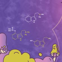
Psychedelics are evolutionarily ancient compounds produced by fungi, plants, and microbes. Humans also synthesize psychedelics. Researchers want to know how and why.

A Chronic Itch: Burrowing Beneath the Skin
Brian S. Kim, MD | Sep 8, 2023 | 9 min read
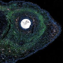
We have barely scratched the surface of itch science and what it indicates about our health.
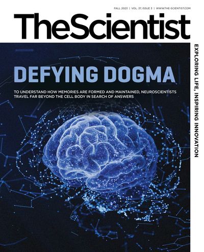
Defying Dogma: Decentralized Translation in Neurons
Danielle Gerhard, PhD | Sep 8, 2023 | 10+ min read
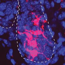
To understand how memories are formed and maintained, neuroscientists travel far beyond the cell body in search of answers.

What Can ChatGPT-like Language Models Tell Us About the Brain?
Natalia Mesa, PhD | Sep 8, 2023 | 8 min read
A renaissance in natural language modeling may help researchers explore how the brain extracts and organizes meaning.
Speaking of Science
What’s the Transmission?
What’s the Transmission?
Put on your thinking cap, and take on this fun challenge.
Editorial
Onward and Upward!
Onward and Upward!
At The Scientist, we are strengthening our roots while reaching for the sky.
Foundations
Magnifying Curiosity with a Pocket Microscope
Magnifying Curiosity with a Pocket Microscope
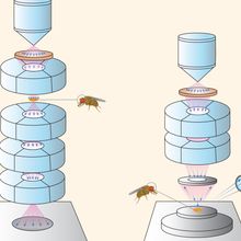
Microscopes were inaccessible to most of the world until Manu Prakash and Jim Cybulski put their engineering prowess to the test.
The Origins and Recent Promise of Nonsense Suppressor tRNAs
The Origins and Recent Promise of Nonsense Suppressor tRNAs
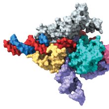
A discovery that goes back to the first studies of translation has become the topic of biotech buzz.
Cell Painting: Exploring the Richness of Biological Images
Cell Painting: Exploring the Richness of Biological Images
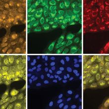
By coloring different organelles simultaneously, cell painting allows scientists to pick up subtle changes in cell function in response to drugs and other perturbations.
The Literature
Mice Heal Themselves in Response to a Common Signaling Molecule
Mice Heal Themselves in Response to a Common Signaling Molecule
A newly discovered way to induce scarless healing in mice depends on a highly conserved signaling pathway that is also present in humans.
New Epigenetic Clocks May
Confirm Extreme Age
New Epigenetic Clocks May
Confirm Extreme Age
How will a new version of epigenetic clocks aimed at validating the age of people older than 100 years of age balance accuracy and anonymity?
CRISPR-like Abilities in Eukaryotic Proteins
CRISPR-like Abilities in Eukaryotic Proteins
Two groups independently discovered that Fanzor proteins in eukaryotic organisms are CRISPR’s genome-editing cousins.
Hair Turns Gray Due to Stuck Stem Cells
Hair Turns Gray Due to Stuck Stem Cells
Hair-coloring stem cells must swing back and forth between their maturity states to give hair its color.
Molecular Signatures of a Broken Heart
Molecular Signatures of a Broken Heart
The transcriptional profiles in the brains of prairie voles changed after a long breakup, revealing a molecular shift that might help them cope with the loss of a partner.
Infographics
Infographic: What a Trip
Infographic: What a Trip
Researchers took a mind-bending trip to understand the connections between psychedelic compounds produced by fungi, plants, and humans.
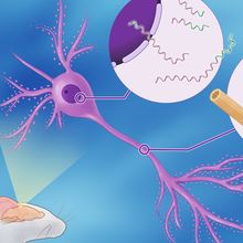
Infographic: Beyond the Nucleus: mRNA Localization in Neurons
Infographic: Beyond the Nucleus: mRNA Localization in Neurons
To support thousands of incoming connections, neurons use sophisticated transportation networks for delivering mRNA to faraway regions.
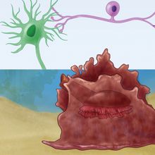
Infographic: Synaptic Plasticity in the Sea Slug
Infographic: Synaptic Plasticity in the Sea Slug
The sea slug has helped scientists in their quest to understand how neurons encode memories.
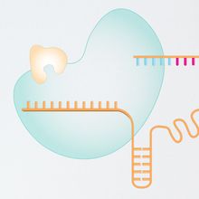
Infographic: How Prime Editing Works
Infographic: How Prime Editing Works
Prime editing is one of the most promising forms of genome editing because it uses only single-stranded DNA breaks.
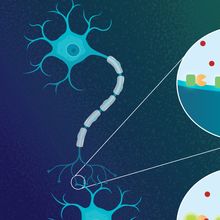
Infographic: How a Glutamate Sensor Tracks Synapses
Infographic: How a Glutamate Sensor Tracks Synapses
A third generation glutamate sensor with a fluorescent readout offers insights into neuronal communication.
Infographic: Drivers of the Expansion of Volume Electron Microscopy
Infographic: Drivers of the Expansion of Volume Electron Microscopy
Technological advancements over the last two decades transformed volume electron microscopy, improving usability, resolution, and throughput.
Profile
A Quest to Drug RNA
A Quest to Drug RNA
Matthew Disney’s idea of small molecules that target RNA once seemed fanciful. Now, even the pharma industry is pursuing it
Lighting Up Diagnostics
Lighting Up Diagnostics
Brought together by a shared interest in synthetic biology and diagnostics, two researchers are transforming how we label biomolecules.
When Microbes Meet the Immune System
When Microbes Meet the Immune System
Timothy Hand leads a research team that explores how maternal immune signals shape the infant intestinal microbiota.
Methods
Prime Editing Comes of Age
Prime Editing Comes of Age
Since the technique was first published in 2019, prime editing has grown with lightning speed, alongside hopes for what it can achieve.
Biosensors Illuminate Talk Between Neurons
Biosensors Illuminate Talk Between Neurons
First developed in 2013, a fluorescent indicator has evolved to enable precise glutamate tracking.
The Expansion of Volume Electron Microscopy
The Expansion of Volume Electron Microscopy
A series of technological advancements for automation and parallel imaging made volume electron microscopy more user friendly while increasing throughput.
