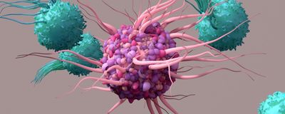
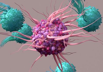
ABOVE: (C) ISTOCK.COM DESIGN CELLS
Vaccination is one of the most effective tools available against infectious disease. Nonetheless, optimizing vaccine efficacy remains a top priority for scientists, especially to better protect high-risk populations. The time of day at which a vaccine is delivered affects response robustness, but how this occurs from a cellular and molecular level is not well understood.1-3 A recent publication in Nature Communications from Annie Curtis’s research group at the Royal College of Surgeons in Ireland linked circadian shifts in dendritic cell (DC) function with oscillations in mitochondrial morphology and metabolism.4 Moreover, Curtis’s team showed that these mechanisms can be directly modulated using small molecule agents—a potentially useful future approach for enhancing vaccine responses.
Most, if not all, biological functions in humans are affected by the circadian clock—stable oscillations in gene expression across a 24-hour cycle regulated by a set of conserved, self-sustaining transcriptional-translational feedback loops.1 Scientists have found that circadian rhythms affect critical immune cell functions, including cytokine production, cell trafficking, and T and B cell activity.1,2 However, the underlying mechanisms driving these time-of-day differences in immune activity—and in particular, the effect of circadian signaling on dendritic cells (DCs)—remained unclear.2
To activate T cells, DCs take up, process, and present antigens as part of major histocompatibility complexes (MHCs) that interact with T cell receptors. Therefore, Curtis and her team set out to examine the effect of circadian timing on DC antigen uptake, processing, and presentation. First, they confirmed the presence of circadian cycling in DCs by looking at the expression of molecular clock genes. Next, they treated two populations of DCs—one with and one without circadian disruption—with the antigen ovalbumin, and found that DCs with intact circadian signaling processed ovalbumin in an oscillating rhythm with a 24-hour period. However, the disrupted population had low levels of antigen processing and no discernable rhythm.
Having clearly established that DC antigen processing was regulated by a circadian cycle, Curtis and company next sought reasons for this phenomenon. Mitochondrial metabolism was a reasonable starting point because it is both a key regulator of DC function and is controlled by the molecular clock. Here, the researchers found a familiar pattern: DCs with intact circadian signaling displayed an oscillating rhythmic phenotype with a 24-hour period, while circadian-disrupted DCs showed no rhythmicity and consistently lower metabolic outputs. Mitochondrial morphology—which directly inf luences mitochondrial metabolism—also changed in a circadian-driven oscillating manner. Specifically, mitochondria in DCs with intact circadian signaling toggled between fused and fragmented phenotypes roughly every 12 hours, while those in circadian-disrupted DCs were predominantly fragmented at all examined timepoints.
Next, Curtis’s team found that directly inhibiting mitochondrial metabolism reduced antigen processing in both intact and disrupted DCs, resulting in significantly lower T cell activity. Disrupting mitochondrial morphological oscillations also affected antigen processing. However, promoting mitochondrial fusion boosted antigen processing during the troughs of the circadian cycle (when mitochondria presented a fragmented phenotype) in molecular clock-intact DCs and rescued the defective antigen processing previously observed in molecular clock-disrupted DCs. Finally, the research team found a critical role for calcium in regulating mitochondrial metabolism and morphology. Not only did mitochondrial calcium localization follow a circadian rhythm in clock-intact DCs (but not clock-disrupted ones) that was mediated by the mitochondrial calcium uniporter, but increasing both mitochondrial fusion and calcium uptake resulted in greater levels of antigen processing than promoting either by itself.
Collectively, these findings indicate that mitochondrial metabolism is a key regulator of antigen processing in DCs and subsequent T cell activation, and that circadian oscillations in mitochondrial morphology driven by calcium levels are mechanistically linked to DC antigen processing. By comprehensively elucidating this process, Curtis and her team not only provided proof of concept that these mechanisms can be modulated using small molecule agents, but also present numerous potential targets for said modulation worthy of future investigation.
References
- Otasowie CO, et al. Chronovaccination: Harnessing circadian rhythms to optimize immunisation strategies. Front Immunol. 2022;13:977525.
- Long JE, et al. Morning vaccination enhances antibody response over afternoon vaccination: a clusterrandomised trial. Vaccine. 2016;34:2679-85.
- de Bree LCJ, et al. Circadian rhythm influences induction of trained immunity by BCG vaccination. J Clin Invest. 2020;130:5603-17.
- Cervantes-Silva MP, et al. The circadian clock influences T cell responses to vaccination by regulating dendritic cell antigen processing. Nat Commun. 2022;13:7217.


