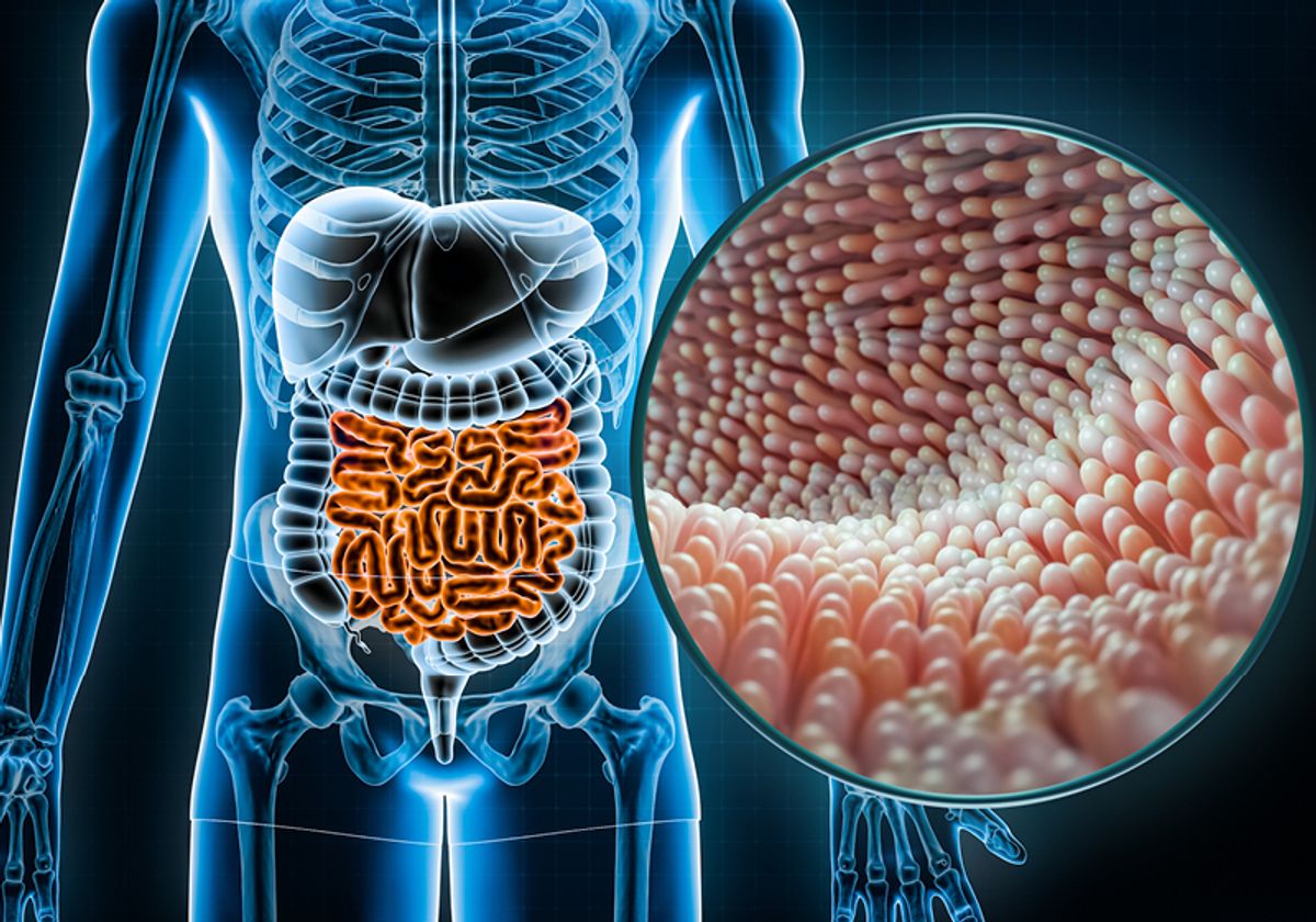
Inflammatory bowel diseases (IBDs) such as ulcerative colitis and Crohn’s disease are syndromes caused by genetic and environmental factors that disrupt the intestinal immune responses.1 Scientists continue to search for more effective and less harmful IBD treatments, which remain elusive in part because current model systems fail to replicate key mechanisms of the epithelial cell monolayer that covers the intestinal surface.1 To address this research gap, researchers turn to new preclinical IBD models to identify and investigate clinically relevant therapeutic targets and biomarkers.1
Models of Inflammatory Bowel Disease
In vivo, the intestinal epithelium interacts with the neighboring environment through complex crosstalk, which involves components such as the immune system, tissue microenvironment, and gut microbes. Preclinical models are simplified tools that help researchers study these complex interactions in a controlled manner and prioritize relevant biochemical pathways when developing IBD therapies. These models include humanized stem cell-derived in vitro systems that recreate the epithelial cell layer makeup and morphology.2
Towards this end, scientists increasingly employ monolayer systems. These cell culture systems are made of a single layer of stem cells, which researchers grow on scaffolds, such as membranes or hydrogels. The cells attach to the scaffold, proliferate, and differentiate into specialized intestinal cell types that recapitulate the composition of the human gut epithelium.2
Different scaffolds allow researchers to finetune their monolayer systems and investigate unique properties of the human intestinal tract. Monolayer systems cultured on porous membranes replicate both the luminal and basal sides of the epithelial cell monolayer. Unlike 3D model systems, monolayers also include epithelial-adjacent fluid reservoirs for researchers to examine external immune cues present in the intestine, such as microorganisms and bioactive compounds. Scientists can turn to monolayer models for simplicity, scalability, and compatibility with multiple assay readouts.2
Monolayer System Readouts
Cytotoxicity
Toxicity is a key consideration when developing new therapies, and the intestinal epithelium is a crucial interface for drug absorption and metabolism in vivo. Because of this, researchers embarking on IBD drug discovery journeys must ensure that therapeutic candidates do not harm the already vulnerable intestinal epithelium. Monolayer systems provide the opportunity to monitor drug toxicity in vitro, allowing researchers to rapidly quantify compound safety and triage unsuitable candidates early in drug discovery.2
Barrier integrity
Healthy intestinal barriers are essential for protection against harmful leakage between the gut lumen and adjacent tissues.3 Scientists study barrier breakdown as an initiating event in IBD. The major barrier cells in the small intestine and colon are epithelial cells, and dysfunction of these cells is of high interest to researchers investigating barrier breakdown.1 Because monolayers embody the characteristic luminal/basal morphology of the in vivo intestinal epithelium, researchers can use these systems to decipher how the intestine monitors its microenvironment for barrier defects and communicates with the immune system.3 Monolayer models typically form a tight layer that supports physiological assays of barrier functions such as ion transport, and barrier integrity such as transepithelial electrical resistance (TEER).2
Inflammatory cytokine production
Researchers can also use these systems to measure the barrier-mediated immune response.2 In vivo, the intestines play a central role in immune system maturation and function; barrier epithelial cells are first responders to IBD inflammation. In response to active immune cells that produce interferon gamma (IFN-γ) and tumor necrosis factor (TNF-α), epithelial cells secrete immune factors, including inflammatory cytokines such as interleukin 8 (IL-8).2,3 Because they mimic the natural barrier morphology with relevant cell types and scaffolds, epithelial monolayer systems support physiological immune assays, including those that measure chemokine inflammatory cytokine synthesis and secretion.2
Monolayer Models for Inflammation-Focused Drug Discovery
Monolayer models hold immense promise as physiologic systems for drug discovery, providing a readout for barrier integrity and allowing scientists to reproduce complex intestinal and immune interactions. The InflammaScreen™ Services from Altis Biosystems are a suite of readout assays specifically designed to accelerate drug screening and improve the human relevance of gastrointestinal inflammation focused drug discovery.4 This suite of assays uses the Altis Biosystem RepliGut® Planar Transverse Colon model system, enabling researchers to rapidly test compounds, compare candidates, and triage ineffective molecules in an inflammatory epithelial monolayer system. The tissue is monitored for barrier integrity using TEER and evaluated for quantitative cytotoxicity and IL-8 release. Researchers can rely on these assays for rapid and clinically relevant insights into intestinal inflammation.
References
- Pizarro TT, et al. Challenges in IBD research: Preclinical human IBD mechanisms. Inflamm Bowel Dis. 2019;25(Suppl 2):S5-S12.
- Dutton JS, et al. Primary cell-derived intestinal models: Recapitulating physiology. Trends Biotechnol. 2019;37(7):744-60.
- Wang Y, et al. Analysis of interleukin 8 secretion by a stem-cell-derived human-intestinal-epithelial-monolayer platform. Anal Chem. 2018;90(19):11523-30.
- InflammaScreen™ Services. Altis Biosystems. Accessed September 15, 2023.


