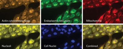
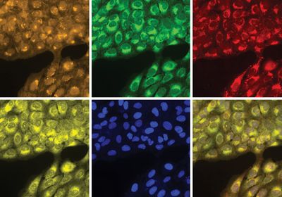
ABOVE: In the cell painting assay, researchers use fluorescent dyes to label different organelles. Broad Institute Center for the Development of Therapeutics
Scientists have been peeking through microscopes and taking pictures of cells for decades. The methods available for visualizing highly complex cells have changed over time, moving from simpler morphology-focused imaging to single-cell specific fluorescence microscopy. Despite these improving approaches, there is a vast amount of information left unexplored in cells, like hidden treasures at the ends of breadcrumb trails.
Anne Carpenter, a cell and computational biologist at the Broad Institute, wanted to explore this vastly untouched world. “As a cell biologist, I felt that labeling the major organelles would probably give us the broadest readout of how the cells were experiencing their environments,” she said.
Changes in the physical characteristics of major organelles can provide great insight into the cells’ states. For instance, in myopathy conditions such as Duchenne muscular dystrophy, mitochondria in muscle and skin cells swell and show reduced matrix density.1
In collaboration with fellow scientists, Carpenter started developing an assay to paint cellular organelles and allow researchers to uncover subtle changes in them. The result was the cell painting assay, which the team introduced in 2013.2
It was a phrase we only used informally in the lab. At some point, we thought about adding 30 dyes at once to the cells and calling it “cell graffiti.”
—Anne Carpenter, Broad Institute
Since Carpenter and her colleagues wanted to paint the organelles simultaneously, the first key step was to choose the right combination of organelle-specific dyes to highlight as many features of the organelles as possible. They came up with a mix of six stains that are distinct from each other as they target specific parts of the cellular structures. As an example, the mitochondria dye binds to the organelle’s membrane, whereas the nuclei stain binds to adenine-thymine pairs in DNA.
As opposed to antibodies that label specific molecules inside the cells, cell painting dyes provide a less biased way of looking at cellular changes since they stain the organelles as a whole, revealing hundreds to thousands of different morphological features such as textural patterns, sizes, and shapes. To create a profile of the cells, scientists then combine all of the features from the different organelles that are imaged and analyzed using cell imaging software such as CellProfiler, another of Carpenter’s creations. The term profiling, Carpenter explained, describes the process of extracting a large set of morphological features from cell images and letting the patterns that arise lead researchers to discoveries.
Early on, the researchers viewed this approach as painting, but they didn’t officially name the assay “cell painting” until later on.3 “It was a phrase we only used informally in the lab. At some point, we thought about adding 30 dyes at once to the cells and calling it ‘cell graffiti,’” she recalled. Eventually, the team pulled back from this more complex approach to keep the assay easy to use and interpret.
If cells with the disease look differently, you can potentially do a drug screening to identify drugs that make the diseased state cells look healthier.
—Anne Carpenter, Broad Institute
“Now that you have a high dimension representation of the state of the cell, any problem that needs to group things together based on biological states is available for being solved,” said Shantanu Singh, a computer scientist who leads a research team with Carpenter at the Broad Institute.
One of these applications is predicting the mechanisms of action of compounds, a process that can be time consuming, especially when evaluating the activity of new molecules, said Slava Ziegler, a biochemist at the Max Planck Institute of Molecular Physiology who uses the cell painting assay in her research. “The cell painting assay is more unbiased because it still focuses on a certain feature, which is morphology of the cells, but not on a certain process, like a signaling pathway,” she explained. By using it, “we thought we would get broad coverage of the bioactivity of all of these compounds synthesized in house.”
As diseases might alter the morphology of organelles, as in the case Duchenne muscular dystrophy, researchers can also use the cell painting assay to identify profiles associated with a disease and compare those to the profiles of healthy cells. “If cells with the disease look differently, you can potentially do a drug screening to identify drugs that make the diseased state cells look healthier,” Carpenter said.
Observing cells through microscopes is a powerful way to look at life on a small scale, Singh explained, and the cell painting assay reveals cellular features that might not have been obvious with other methods. From a qualitative description to the measurement of a few and now thousands of features, Carpenter believes that cell painting is changing the way researchers look at these highly complex biological snapshots. “It is pretty exciting for researchers to use images as a data type as opposed to just confirming what they see by eye,” she said.
References
- Pellegrini C, et al. Melanocytes—a novel tool to study mitochondrial dysfunction in Duchenne muscular dystrophy. J Cell Physiol. 2013;228(6):1323-1331.
- Gustafsdottir SM, et al. Multiplex cytological profiling assay to measure diverse cellular states. PLoS One. 2013;8(12):e80999.
- Bray MA, et al. Cell painting, a high-content image-based assay for morphological profiling using multiplexed fluorescent dyes. Nat Protoc. 2016;11(9):1757-1774.


