Cartoon showing someone on stepping stones to success

Failing to Succeed

We must celebrate scientific failures not as precedents to anticipatory successes but for what they are: valiant efforts that didn’t work out. 

In 1928, Alexander Fleming noticed a mold infiltrating his Staphylococcus bacteria culture plates. Fleming observed that the intruder inhibited bacterial growth and eventually found the causative antibiotic: penicillin. 

This story of a serendipitous groundbreaking discovery from a mishap is an inspiration to all scientists. It offers hope that even if an experiment fails, something extraordinary may come out of it. While everyone needs this motivation to keep going on rough days, such success stories set unrealistic expectations of a strong comeback. A more realistic scenario is that researchers lose time, samples, and effort, with no compensatory gains. 

Survivorship bias runs rampant in science. As journalists, we are privileged to cover cutting edge research and inform the community about seminal updates in life science. We report on the best publications and shine a spotlight on the study authors for their wins. These articles keep researchers motivated, informed, and excited about science, but reading only about successes makes them feel like the norm, even if these big advances are rare. This feeds the monster of imposter syndrome in an already frail “publish or perish” academic ecosystem wherein failure may feel shameful. 

So, what can we do about it? Failing is normal, but we need to normalize talking about it. Sometimes scientists fail and then eventually succeed, and sometimes they simply fail to succeed. We want to normalize both scenarios. As a step in this direction, I am thrilled to introduce our new column, “Epic Fail,” which will provide an outlet for scientists to share their failures. In our first column, Gaurav Ghag from Gilead Sciences shared how he handled the disastrous realization that he had replicated a calculation error in an experiment over the course of four years during his graduate work. 

Whether a doomed experiment became a stepping stone to success, remained a funny anecdote, or served as a lesson for personal growth, we want to hear about it! We hope to create a fail-safe space for scientists and foster camaraderie through commiseration. We look forward to your best failure stories.

Send Feedback

Image of unfolded protein

My Protein Didn't Fold and Neither Did I

When Gaurav Ghag realized that he had replicated a calculation error in every experiment during four years of his graduate research, he initially thought that his career had unraveled with his protein.  

In 2016, I was a fourth-year graduate student at the University of Southern Mississippi. I studied the structure of granulins, which are cysteine rich proteins implicated in neurodegenerative diseases. To verify protein folding, I conducted an assay so often that I created a templated, ready-to-use protocol for it.

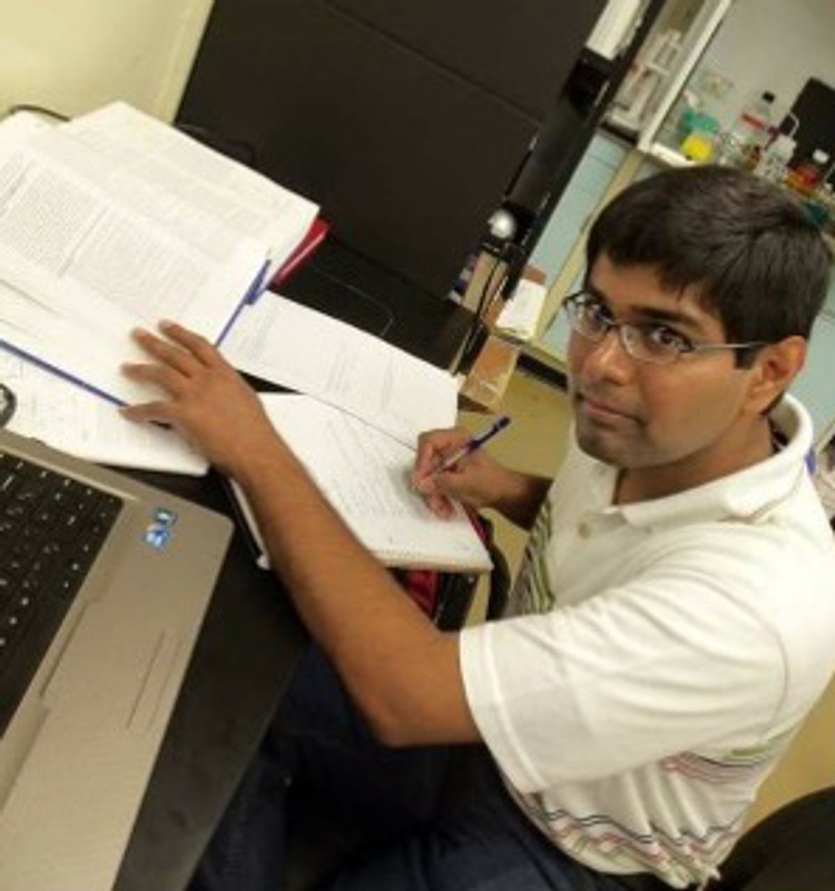
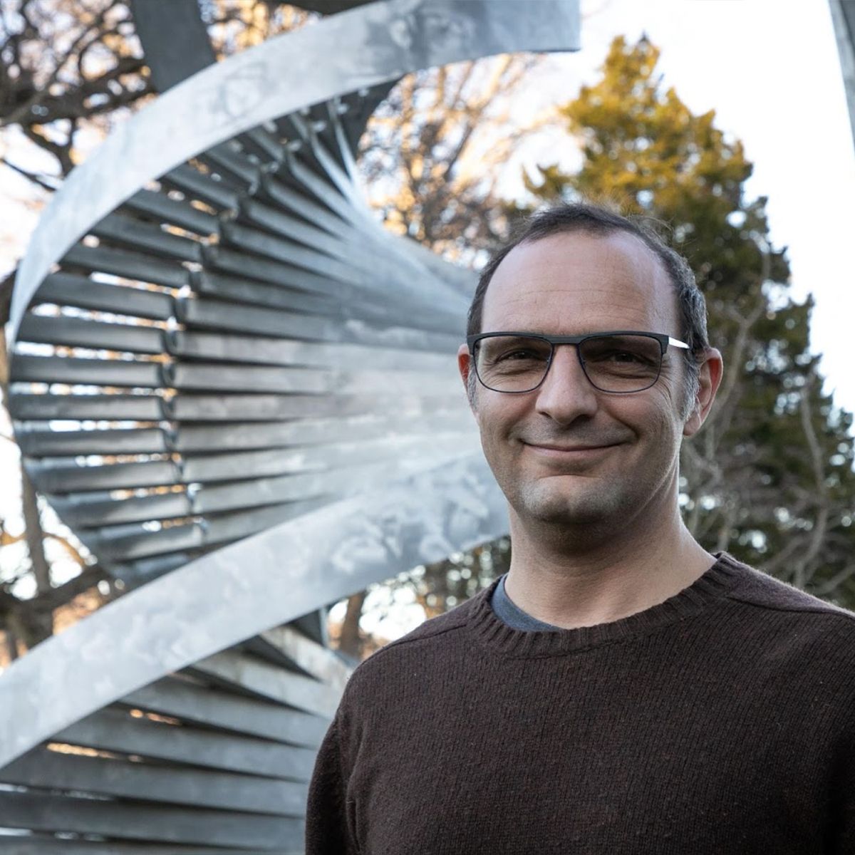
          Image of Gaurav Ghag
Gaurav Ghag conducted his graduate research at the University of Southern Mississippi. He is now a senior manager of analytical operations at Gilead Sciences.
Michael Samel

As I inched towards the end of graduate school, I was excited. I had fantastic data on my folded protein, and a high impact journal publication seemed within reach. 

One day, I tested an alternative method to assess granulin folding, and weirdly, it indicated that the protein was unfolded. Perplexed, my advisor, Vijayaraghavan Rangachari, and I walked through every step of the experiment until it struck me: I had made a decimal point calculation error in my templated protocol! I realized with dismay that I had replicated this mistake in every experiment. I had been working with unfolded protein all along. 

Shocked, I exited my advisor’s office, poured myself some coffee, and just sat outside for hours in disbelief and disappointment. I had no time to start a new project or to repeat my experiments. What was I going to do?

Once I calmed down the next day, I decided that it wasn't over. I still had highly reproducible (unfolded) protein data that might be worth something. With the support of my advisor, I managed to publish the data.1 It didn't end up being the high impact factor paper that I dreamed of, but we managed to put a positive spin on a bad situation. 

I view that incident as the biggest learning moment of my life. First, the devil is in the details, so I always have someone else double check critical work now. Second, I remind myself that mistakes happen, but resilience helps overcome them. 

Although I have moved on since, I often remember a thought from that day: "Cysteines, thou art heartless beasts!"

This interview has been edited for length and clarity.

Reference

  1. Ghag G, et al. Protein Eng Des Sel. 2016;29(5):177-186.

Submit Your Story

ADVERTISEMENT
Image of T cell binding to site

Multiple Targets, Infinite Possibilities

Multispecific antibodies are rising stars in the field of antibody therapeutics, offering better specificity, targeting ability, and therapeutic effects than traditional monoclonal antibodies.

          Cartoon-style illustration of a bispecific antibody bound to a T cell through one binding site and a tumor cell through a second binding site
Multispecific antibodies bind to two or more different epitopes, enabling physically linked multitargeting with a single therapeutic.
SINO BIOLOGICAL

Antibody-based therapeutics have revolutionized treatment options for many indications, including cancer, autoimmune and metabolic conditions, and infectious diseases. Although monoclonal antibodies (mAbs) currently account for the majority of biologics approved annually by the FDA,1 mAbs bind to only one target by design. The therapeutic success of mAbs has inspired researchers to push antibody therapeutic technologies toward multifunctionality, turning to bispecific, trispecific, and tetraspecific antibody design and development for increased specificity and efficacy while reducing side effects.2

Scientists have created multispecific antibodies with the ability to bind two or more different antigens or two or more different epitopes on the same antigen for novel applications, benefitting from physically linked multitargeting in a single therapeutic. 

Compared to monoclonal therapeutics, researchers face additional challenges during the different stages of multispecific antibody discovery and development.2 Scientists address increased complexity of multitargeting with high quality target proteins, such as CD3, multipass transmembrane, and immune checkpoint proteins from Sino Biological. High quality targets accelerate translational multispecific antibody research for clinical applications and support researchers through each stage of discovery and development.

References

  1. de la Torre BG, Albericio F. Molecules. 2022;27(3):1075.
  2. Labrijn AF, et al. Nat Rev Drug Discov. 2019;18(8):585-608.
Learn More
Tumor microenvironment concept with cancer cells

Cancers Protect Themselves Against Their Own Mutations

Tumors overexpress certain genes to survive a growing pile of harmful mutations, a trait that scientists could exploit to target with drugs.

Most cancerous tumors accumulate thousands of potentially protein-damaging mutations over time, yet they mysteriously continue to thrive.1 Now, a new computational study helps explain how that is possible: Tumors with a large number of mutations upregulate genes that minimize misfolded proteins to protect them from their own mutations.2

“[These mutations] very likely accrue over decades, and the cancer cells need a way to cope,” said computational biologist Christina Curtis from Stanford University, who coauthored the study. 

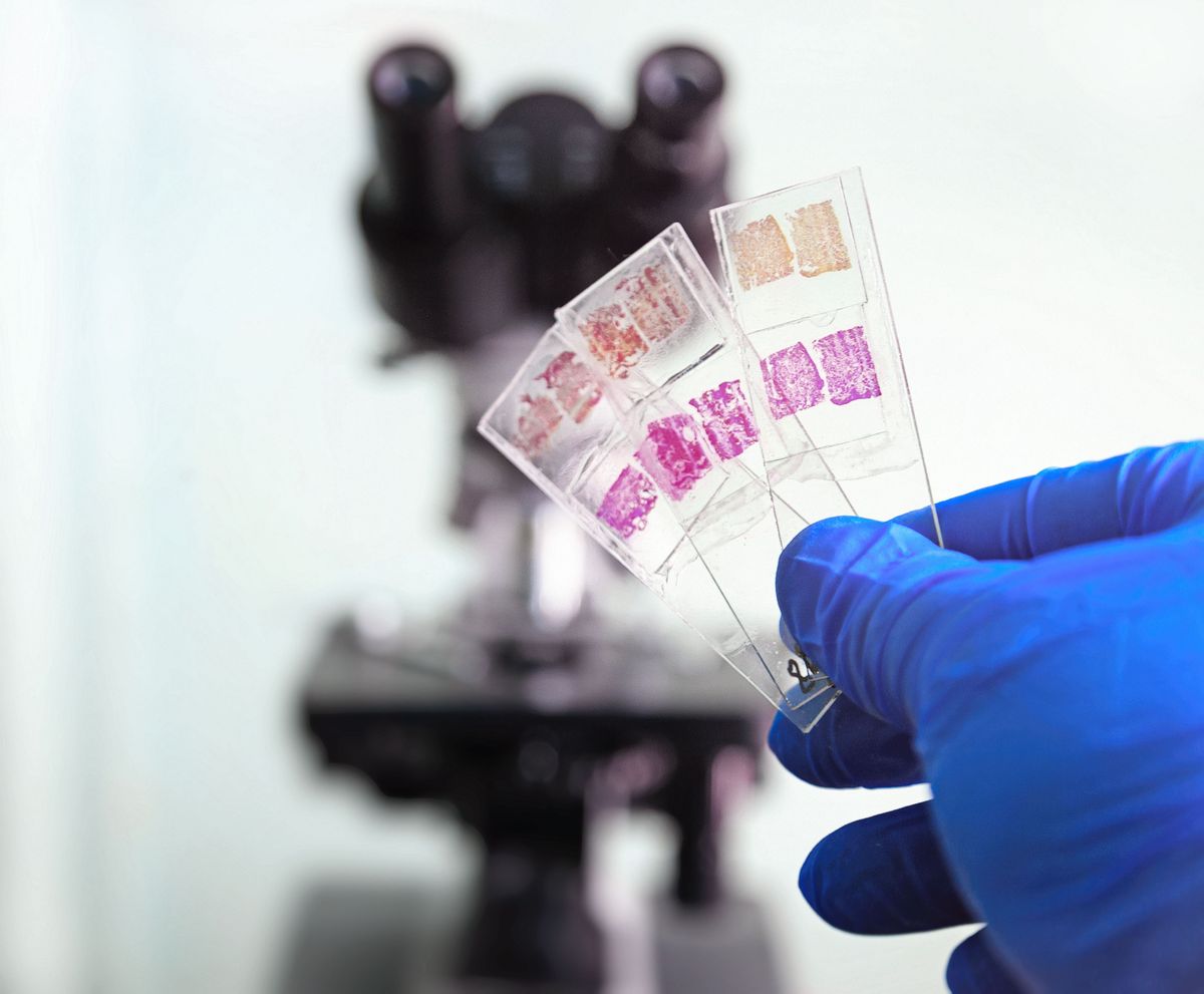
     A gloved hand holding three glass slides with stained tissue samples in front of a microscope.
By analyzing the gene expression of more than 10,300 human cancer tumors, Christina Curtis and her team at Stanford University discovered that cancers protect themselves from their own harmful mutations.
© ISTOCK.COM, Kostafly

To reveal that coping mechanism, Curtis and her team explored the gene expression of nearly 10,300 human tumors across 33 cancer types catalogued in the Cancer Genome Atlas database.3 They found consistent upregulation of chaperone proteins and the proteasome, which respectively prevent and degrade misfolded proteins. 

Next, the researchers validated their findings using cell line data from the Cancer Cell Line Encyclopedia.4 The cell lines showed similar expression patterns, and when the team calculated the effect of knocking down the upregulated genes, higher mutational loads correlated with reduced cell viability. These results suggest that the gene upregulation protects tumors.

For Curtis, this discovery signals a general vulnerability in many tumors that could be exploited, for example by using chaperone and proteasome inhibitors. Scientists developed such drugs decades ago, but this new information might help target them to the tumors that will be most vulnerable.

Cancer geneticist Ekta Khurana from Weill Cornell Medicine, who was not involved in the study, said that this was an exciting finding that looked beyond the mutations that help cancers grow. “[It] beautifully shows that broadening our perspective can lead to not just insights about how cancers evolve, but actual therapeutic opportunities.”

References

  1. Tilk S, et al. Elife, 2022; 11:e67790.
  2. Tilk S, et al. Elife, 2023; 12:RP87301.
  3. Ellrott K, et al. Cell Syst, 2018; 6.3: 271-281
  4. Barretina J, et al. Nature, 2012; 483: 603-607.
ADVERTISEMENT
Digital illustration of neurons

Captivated by the Great Expanse of Neurons

According to Erin Schuman, science driven by fascination rather than tools will guide new discoveries.

Erin Schuman, a neurobiologist at the Max Planck Institute for Brain Research, studies how information is processed and stored in neurons. Schuman is the corecipient of the 2023 Brain Prize for her groundbreaking discoveries on local translation and synaptic plasticity.

          Image of Erin Schuman
Erin Schuman, a neurobiologist at the Max Planck Institute for Brain Research, studies the formation and maintenance of synaptic connections in the brain.
Gilles Laurent

What drew you to study cell biology in neuroscience?

What captivates me is how the neuron functions as an individual cell and how its morphology changes its biology. Most cells in the body are spherical, but neurons are unique. Around 80% of a neuron’s volume is in its extensive protrusions: axons and dendrites. The neuron has engineered a way for protein synthesis machinery to work beyond its cell body and into its expansive volume to preserve the functional integrity of its 10,000 synapses. That is something special. 

What major challenges do neuroscientists face today?

Neuroscience is much too driven by the availability of tools and model systems. Instead of people saying, “what is a question I really want to ask?”, they ask, “what is the technique that I want to use?” They may do that consciously or subconsciously, but I find that science driven in that way is not as interesting and is less likely to lead to new discoveries. We need more original thinking that is not driven by the availability of tools. 

What advice would you give to early career researchers? 

Find a question that completely captivates you—one that you can muse about endlessly. Choose one that is not something that can be easily answered; explore a deep conceptual issue that you find very exciting personally because that will lead to excitement in your experiments. It will motivate you to stay in science and work on your project, even in the face of adversity. The things that captivate you are more likely to be where you are going to make an impact.

This interview has been condensed and edited for clarity. 

Image of ancestor with hair all over body

Why Don’t Humans Have Fur?

Humans are often referred to as “hairless apes.” But how did this trait evolve given that fur provides significant advantages to many mammals?

          Headshot of Nina Jablonski
Nina Jablonski, a biological anthropologist and paleobiologist, studies primate and human evolution at Pennsylvania State University. 
Nina Jablonski

Luscious fur coats insulate many animals from the cold and protect them from sunlight, insects, and sharp objects in their environments. Yet, somehow humans evolved to be relatively hairless. While this may appear to be a case of selection against a highly desirable trait, Nina Jablonski, who studies the evolution of human skin and skin pigmentation at Pennsylvania State University, said that our relative hairlessness arose just like other traits did: it offered evolutionary advantages.

The origins of human hairlessness began nearly two million years ago, driven by environmental changes in locations where human ancestors lived. As wooded landscapes in equatorial Africa gave way to open grassland areas, human ancestors had to spend more time outdoors to find food and water. For walking and running long distances, early members of the genus Homo developed a modern human skeleton with long legs and shorter arms. “Around this time, humans lost most of their body hair,” said Jablonski. 

Shedding body hair was a key adaptation since, unlike most other mammals, primates lack a key mechanism for cooling the blood around the brain when it’s hot outside or after exercise. This means that the temperature of the brain increases when the body heats up, which can affect brain functions. Evolution of human hairlessness was accompanied by the development of more sweat glands and darker skin pigmentation. Sweat glands helped them dissipate heat from the skin more effectively, while darker skin pigmentation protected their mostly hairless skin from the damaging effects of solar radiation.

According to Jablonski, the idea of whole body cooling and heating, or thermoregulation, seems like the most likely explanation for human hairlessness based on physical evidence and our knowledge of comparative anatomy and physiology. “We were shooting in the dark decades ago. Now, we can be much, much clearer on what the likely courses of evolution were,” she said. 

What makes you curious? Submit a question for us to answer in future “Just Curious” columns. 

Submit a Question

ADVERTISEMENT

A Brain MicroRNA Curbs Anxiety

Upregulation of a specific microRNA in the brain lessened anxiety and reduced the expression of stress-related genes in mice. 

Stress is a known risk factor for anxiety disorders, which affected more than 300 million people worldwide in 2019.1 While antianxiety medications exist, these drugs do not work for everyone. Valentina Mosienko, a neuroscientist at the University of Bristol, strongly believes that we need better treatments for anxiety, but the first step is to understand its molecular basis.  

With that in mind, she and her team explored microRNA since altered levels link to psychiatric disorders.2 How microRNA regulates stress in the brain is largely unknown. In a recent Nature Communications publication, Mosienko and her colleagues reported the role of a specific microRNA, miR-483-5p, in stress-induced anxiety in mice.3 

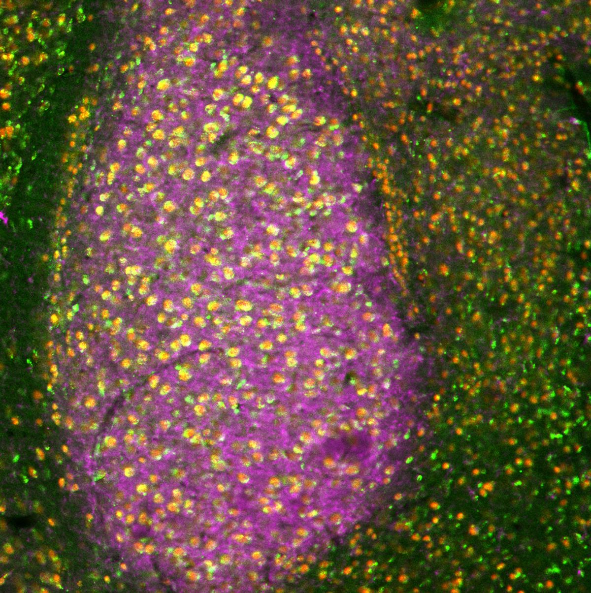
          The image shows a brain section of the mouse amygdala. Using fluorescent markers, the expression of synapses is shown in purple, while neurons are shown as red dots and the microRNA miR-483-5p is shown as green dots.
Mosienko’s team performed fluorescence in situ hybridization on mouse amygdala such as the one pictured above. The expression of miR-483-5p is shown in green. Red indicates the expression of a neuronal marker, while the purple color represents the expression of a synapse marker. 
Mariusz Mucha

Mosienko and her colleagues focused on the amygdala, a brain region implicated in anxiety and control of the stress response. The team stressed mice by restricting their movements and harvested their amygdalae to detect microRNA expression changes. Stress increased the expression of a handful of microRNAs, among which miR-483-5p was the most upregulated. The team also found that miR-483-5p downregulated stress-related genes, particularly Pgap2, in neuronal cell cultures exposed to a synthetic stress hormone. 

To test the effects of miR-483-5p on anxiety-related behaviors, the researchers injected the microRNA into amygdalae in mice and found that it reduced anxiety after stress. Injecting the mouse amygdalae with both the microRNA and an miR-483-5p-resistant Pgap2 reverted this effect as miR-483-5p was unable to act on its Pgap2 target. 

“This microRNA is good,” said Gerhard Schratt, a professor of systems neuroscience at the Swiss Federal Institute who was not involved in the research. “It seems to be more a counter response of the cells in order to dampen the stress and  anxiety.” 

Mosienko hopes that these findings will pave the way for better anxiety treatments as the brain area and molecular pathways the team investigated are similar to those in humans. “We could try to create a molecule that might enhance this pathway to increase our resilience to stress or start thinking about a pharmacological agent that might help people feel less anxious,” she said.

References

  1. World Health Organization. Mental disorders. 2022.
  2. Issler O, Chen A. Nat Rev Neurosci. 2015;16(4):201-212.
  3. Mucha M, et al. Nat Commun. 2023;14(1):2134.
Image representing states of consciousness

High Time: The Roles of Endogenous Psychedelics

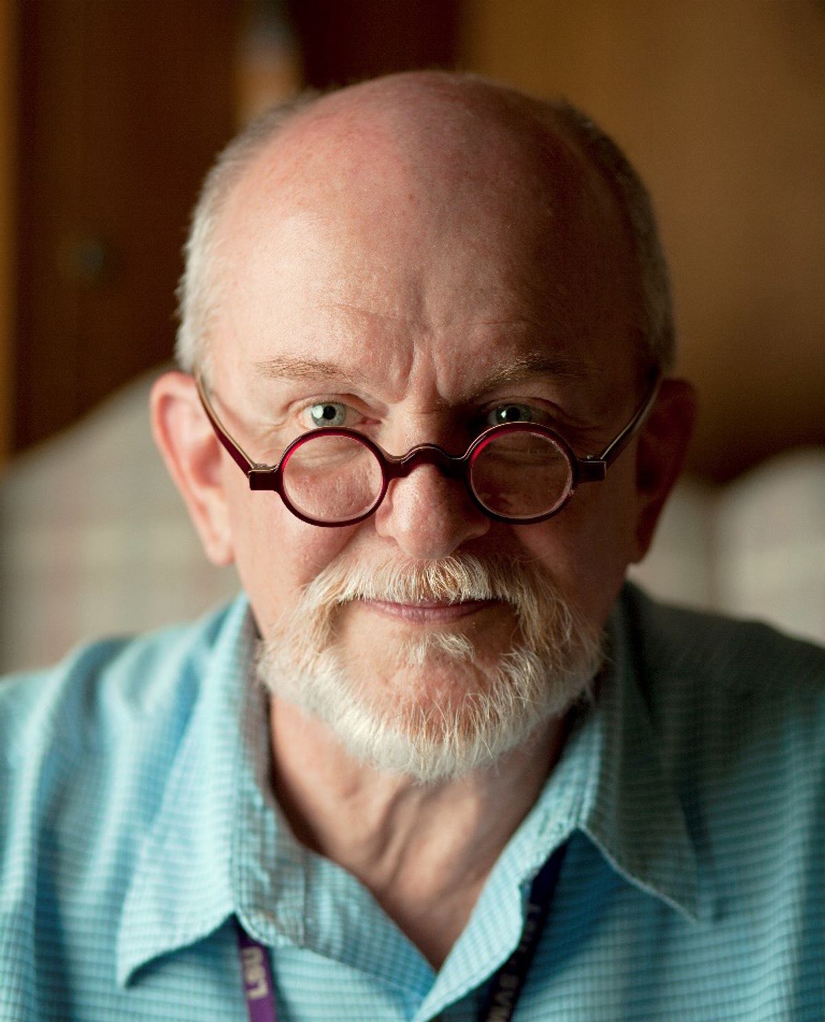
Steven Barker is on a forty-seven-year-long journey to understand the mind-blowing science of psychedelics.

Steven Barker is a professor emeritus at Louisiana State University. His research career spans almost five decades, during which he studied how hallucinogens work, whether humans produce psychedelic compounds, and what role they may play in neuroprotection, psychiatric disorders, perception, creativity, imagination, dream states, and near-death experiences.

          Portrait of man with a white goatee wearing an aquamarine shirt and burgundy spectacles.
Researchers study the extraordinary effects of hallucinogenic drugs on humans, but Steven Barker is fascinated by the roles that endogenous hallucinogens play in ordinary human experiences.
steven barker

What interests you about psychedelics research?

People find psychedelics interesting because of the hallucinations they produce, but they also play physiological roles at much lower concentrations. Psychedelics help grow, maintain, and repair neurons in the brain. Endogenous psychedelics produced by the human body may also protect neurons from hypoxia. 

Dimethyltryptamine (DMT) is a plant extract that elicits powerful psychedelic experiences. Humans and animals also produce DMT in small quantities. We showed that inducing hypoxia in rats increased endogenous DMT levels in the brain and that inducing cardiac arrest enhanced these levels further.1 In fact, elevated endogenous DMT may underlie hallucinations during near-death experiences. Endogenous psychedelics may also be involved in natural hallucinatory-like states such as creativity, imagination, and dreaming. 

How has the psychedelics field evolved?

We lost about twenty years when hallucinogenics research was illegal beginning in the 1960s. Rick Strassman—a clinical associate professor of psychiatry at the University of New Mexico School of Medicine—broke the ice in the 1990s when he started DMT research. Since then, scientists have learned that exogenous psychedelics derived from plants and fungi can help treat depression, post-traumatic stress disorder, and addiction. 

We published data on DMT in the pineal gland and visual cortex of the rat brain and found indirect evidence that the enzyme and precursors needed for its biosynthesis, storage, and release are present in the human brain. New information is emerging about the previously unrecognized roles of psychedelics, but their acceptance will take time given the history and myths surrounding hallucinogens. 

This interview has been condensed and edited for clarity.

Reference

  1. Dean J, et al. Sci Rep. 2019;9:9333.
ADVERTISEMENT
Different colorful coral reefs species surrounded by different fishes. ?

A Probiotic to Protect Caribbean Corals

A bacterial strain from healthy corals could slow the progression and prevent transmission of the destructive stony coral tissue loss disease in the wild.

White, lifeless skeletons stand where bright colored corals once existed. That’s the aftermath of stony coral tissue loss disease (SCTLD), a transmissible waterborne disease of unknown cause that has spread across the Caribbean, affecting more than 20 coral species. Antibiotics stop SCTLD progression, but they do not protect corals from reinfection. 

Now Valerie Paul, a marine biologist at the Smithsonian Marine Station at Fort Pierce, and her team have found a unique way to fight off SCTLD: treating diseased corals with bacteria from healthy corals. Her findings, published in Communications Biology, reveal a potential option for protecting coral reefs in the wild.1

The study launched when Paul and her team observed that some corals remained uninfected despite being housed near diseased corals of the same species in their lab aquariums. Intrigued, the team investigated the microbiota of healthy corals and isolated a Pseudoalteromonas strain, McH1-7. When the team cocultured McH1-7 with the bacteria from diseased corals, they observed that McH1-7 killed other bacteria. Next, they sequenced DNA from this strain and found a gene cluster tied to a known antimicrobial, korormicin.

The team next tried treating diseased corals with McH1-7 and found that it stopped SCTLD progression in aquarium experiments. Also, healthy corals pretreated with McH1-7 were not infected by SCTLD when exposed to diseased corals. 

Since this microbe is already part of the coral’s microbiota, it poses a very low risk for treating corals in the wild, said Kimberly Ritchie, a marine biologist at the University of South Carolina Beaufort who was not involved in the research. “We need to come up with an alternative to antibiotics. Since bacteria are some of the big producers of antibiotics, it makes perfect sense to use nature to help control diseases,” she said. 

The next step is to come up with ways to scale up McH1-7 usage in the wild, noted Paul, since it is currently only applicable on a coral-by-coral basis. “The whole problem with the disease is the scale,” Paul said. Since the disease affects thousands of corals, she hopes that they can find a widespread approach for treating many at the same time. 

Reference

  1. Ushijima B, et al. Commun Biol. 2023;6(1):248.

Relevant Models Reflect Real-world Needs

Jie Sun shares how his curiosity, creativity, and motivation to address clinical public health needs steer his research in immunology and infectious disease.

Jie Sun is a professor of Infectious Diseases and International Medicine at the University of Virginia School of Medicine. In their latest work, Sun’s research team identified genetic and pharmacologic pathways that attenuate severe flu or COVID-19 infection and reduce blood glucose levels that spike after viral pneumonia.

In this episode, Deanna MacNeil spoke with Sun to learn more about his philosophy of science, which prioritizes physiologically relevant models of infection to tackle real-world clinical needs with research.

    Portrait of Jie Sun
jie sun
Jie Sun, PhD
University of Virginia School of Medicine

Science Philosophy in a Flash focuses on the people behind the science. This podcast highlights researchers’ unique outlooks on what motivates their pursuit  of science and what it means to be a scientist. 

ADVERTISEMENT

How should we proceed with image-analyzing AI?

Algorithms can now glean ever more molecular and genetic information from images of stained tissue, but some researchers worry that we can’t follow their logic.

Scientists are using AI-based models to scrutinize disease biology like never before, and each model is more revealing than the next. Deep learning algorithms can now find cancerous mutations,1 estimate the mutational burdens of tumors,2 and predict key gene expression3—all based on stained tissue images. However, because most of these models are “black boxes” and independently learn which features connect to disease, their reasoning can be difficult to decode.4 Does that matter and should it worry us? We asked two experts how they feel about the algorithms’ mysterious natures.

     Headshot of Liron Pantanowitz
Liron Pantanowitz is a computational pathologist at the University of Pittsburgh.
The University of Michigan
Liron Pantanowitz
We should be excited but cautious. We are entering a new era of AI where some people worry that we are training algorithms to process data in a black box. Specifically, we are unsure if we are training them appropriately. However, despite their opacity, these models can be very helpful. For example, they can help us make new discoveries in spatial biology, like how tumors respond to immunotherapy in 3D. Deep learning algorithms can analyze spatial parameters and tell us things that would be inaccessible without them. We’ll be able to discover new diseases and responses to drugs that we’ve never been able to see before. That is super exciting.
     Headshot of Luigi Marchionni
Luigi Marchionni is a computational biologist at Weill Cornell Medicine.
Elisabetta Girardi
Luigi Marchionni
We need to be careful. Deep learning algorithms are intrinsically not very interpretable; they might give us the correct answer, but we don't know why. We cannot open the box and look inside. That is the perfect recipe for creating a biased model because it might not be trained on data that represents the population where it will eventually be used. It is like biomarker development, where we might overfit a model to noisy data, so it only works for that data set and not for the next one. But because AI is so powerful, it’s a much bigger problem. We should avoid the hype and spread awareness of the challenges.

These interviews have been condensed and edited for clarity.

References

  1. Chen M, et al. NPJ Precision Oncology. 2020;4(1):14.
  2. Jain MS and Massoud TF. Nat Mach Intell, 2020;2:356-362.
  3. Anand D, et al. J Pathol Inform. 2020;11(1):19.
  4. Lipkova J, et al. Cancer Cell. 2022;40(10):1095-1110.
August 2023 crossword image

It’s Crossword Time

Put on your thinking cap, and take on this fun challenge.


          August 2023 crossword
Click the puzzle for a full-size, interactive version.
Stella Zawistowski
ACROSS

8. Having both male and female characteristics
9. Material used in laboratory spatulas
10. ___ antigen detection test
11. Takes in air
12. Ion that can be pumped by cells against large gradients
14. 0.01 Gy
16. Motif found in proteins of the inner nuclear membrane
18. Deviating from the expected value
21. Carbon dioxide and water from cellular respiration
22. Not glossy
23. Way to quantitatively measure proteins in mixtures: Abbr.
24. Aggregate that may be magmatic or hydrothermal

DOWN

1. Network that includes unmyelinated nerve fibers
2. Mollusk that clings tightly to rocks
3. Container of a plant embryo
4. NASA spaceflight program from 1961 to 1966
5. Condition with a lack of adequate blood supply
6. German astronomer known for his laws of planetary motion
7. Mountain range where Mouflon sheep can be found
13. Bones whose name comes, appropriately, from the Latin for "shoulder"
15. Old method of portable data storage
17. Material between cells, in eukaryotes
19. Like some football kicks
20. Between 90 and 180 degrees
21. Word before "data" or "ocean"
22. Signaling pathway that is activated in many breast cancers

ADVERTISEMENT